International Hepato-Pancreato-Biliary Association
Left lateral sectionectomy [case 12]
Post-op Debrief
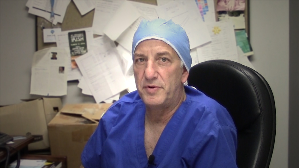
Watch the following videos for the surgeons summaries of the case.
Key points from this case
Neovascularization: Hepatomas typically lead to neovascularization; resulting in arteries, hepatic veins and even portal veins to be larger than usual.
Revising surgical margins: When margins need to be revised, be aware of new potential slowing down moments.
The issues that the surgeons collectively identified preoperatively:
• Uncertain location of tumor
• Possible procedures: Sg2-3 wedge resection, left lateral sectionectomy, left hepatectomy
• Dividing the left hepatic vein
• Sg2-3 left hepatic artery
• Sg3 vein from left hepatic vein
Dr. Gallinger's transcript.
The issues were demonstrated in the intraoperative videos. After the usual preparations, we felt that a midline incision would be adequate. Patient is thin, all of the action of the surgery would be on the left, it was unlikely that we’d have to mobilize the right part of the liver or go anywhere near the vena cava. In which case a midline is faster, less painful, easy and gives us good access. So we made a midline incision and we could immediately see this large tumor which was very exophytic as planned, as predicted. There was no evidence of metastatic disease in the portal lymph nodes, within the liver, or the abdomen. She’d had a prior CT of the chest which was clear so we felt it was operable.
There was very little mobilization because it was so exophytic. In fact, we just literally lifted up the left lateral segment by the tumor kind of like a handle. We eventually decided not to divide the ligamentum venosum which I would normally divide doing a formal left lateral segmentectomy but it looked like the majority of this tumor was actually hanging off of Segment 3 and far enough away from the falciform ligament and the major vessels that we could do a Wedge or a sub-segmentectomy. So we incised the falciform ligament on the left to not go anywhere near the feedback vessels to Segment 4 on the right and went around the pedicle of the Segment 3 and divided it. Initially with a silk tie and then with a clip. There was no replaced left hepatic artery and Segment 3 demarcated quite nicely and we started going through the parenchyma with the goal of at least a centimetre margin which was easy. We incised the capsule of the liver and started dissecting. Unfortunately, either the clip or the tie on that Segment 3 vessel came off, started bleeding. We had to oversew that. We then continued the dissection.
I decided for ease of equipment, save some money, save some time, to use the Kelly crush technique. Which has been proven to be a little bloodier with higher blood loss but probably faster and I think is under-used nowadays, especially in a relatively easy resection where there’s very little risk of loss of control or bleeding. Essentially, there’s lots of ways to do it and everybody has their own little tricks but essentially you use a clamp, we use a Kelly, and you crush the parenchyma and you just need to get a feel for the texture which is different in everybody. If you can guide or gauge the texture through your hands, through your fingers just tensile, or just by feeling it with your fingers, you can actually crush all the parenchyma and preserve the vessels and the bile ducts. Tiny vessels will tear and you can bleed those but large vessels you can spare and when you see them, like you would with any dissection technique, you basically tie them off or clip them or whatever is necessary. That worked very well through the parenchyma.
Towards the end or in the middle, unfortunately there was an arterial bleeder suddenly. Probably Segment 2. It was controlled by pinning that vessel. At that point the specimen had been pretty much removed but we also noticed that the remainder of the left lateral segment was ischemic and would be necrotic and we were going to leave a reasonably large piece of necrotic liver in, which may or may not cause problems. But we felt, since things were going well otherwise, that we should remove the remainder. To complete the operation which otherwise went smoothly, we used the endo-GIA staplers for the large hepatic vein trunk at the back of the liver which was actually quite predictable from the CT scan because she did have quite a large hepatic vein.
The other thing to appreciate is that hepatomas have this habit of grabbing, stealing blood vessels and there’s a lot of neovascularization so arteries are bigger than they normally are, even portal veins, hepatic veins; and that was the case here but it was a pretty easy resection except for the vascular issue that was mentioned.
Dr. Jagannath's transcript
I must highlight in this particular case, very important anatomical planes of the left portal pedicle. The left portal pedicle which comes like that, turns anteriorly at the umbilical point and gives the blood supply to both Segment 2 and 3 on one side and Segment 4 on the other side. Because the Segment 2 and 4b vessels start lower down and go up, you are very prone for injury. Here because of the non-anatomical nature of the resection, we actually ended up in having to perform an anatomical resection because the vascularity has been hit. How do we prevent this kind of an occurrence? One thing is intraoperative ultrasound has to identify what is called the umbilical point or the U-point from which the Segment 2 and 3, two separate pedicles are always supplied. It’s also important to understand that once you cross the falciform, you are unlikely also to hit the Sg4 vessels so this is a very important point for us to understand the blood supply to the left lobe of the liver coming from the portal pedicle at the umbilical point and dividing into the lateral and medial structures. These are very close by and this is something which always happens unless you are very careful about the vessels.
Also, very important for us to understand, along with the portal pedicles are the biliary ducts and unless you see them, transfix them and fix them, these are the patients who are liable for a biliary leak in the postoperative period and one has to be very careful of such occurrences because biliary leak is the one that produces sepsis and the post operative problems because of biliary sepsis are very common.
So coming back to this case, if you see this a second time, classically between the caudate lobe and Segment 2 and 3, is the plane of Arantius’ canal; unless you open this plane, you can then comfortably take the left hepatic vein whether it is intraparenchymal or extraparenchymal, it doesn’t matter and that makes life very easy for completion of a formal hepatectomy. The question then comes on why not a left hemihepatectomy and why a left lateral segmentectomy? This is a decision that was based initially on our preoperative imaging, on the proximity of the tumor to the gallbladder fossa and it is likely that it may be something which is displacing it but yes, if you are planning for a left hemihepatectomy and you still do a left lateral segmentectomy, that is fine but if you do not plan for a left hemihepatectomy, and only try to plan for a non-anatomical resection, and then you convert it into formal resection, that’s when you know you’ll get into problems. In this particular case because there is no cirrhosis, the two splits were done fairly comfortably but this can be a bloody mess and one has to avoid blood loss in such a simple resection as this.
Acknowledgements
Thank you to the HPB Surgeons who contribute their time and expertise. This content is made possible through educational grants from:

![]()
Views and opinions expressed in all videos and module content are those of the individual surgeon and solely intended for surgical education purposes. We do not endorse any product, treatment or therapy.
Corporate Partners
If you are interested in becoming a Corporate Partner of the IHBPA please contact industry@ihpba.org
Find out more


